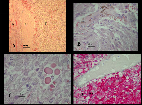
Figure 3.
Histology data. A, low magnification showing the watershed between tumor (T) and normal gland (N), and the tumoral "pseudocapsule"(C). Organoid ( alveolar) arrangement of tumor cells with nests or cords of poligonal eosinophilic tumor cells in a highly vascularised stroma.B, tumor cells showing finely granular, pinkish-brown, delicate cytoplasm, ovoid nuclei with small regular nucleoli. Haemosiderin from small intratumor haemorrages is shown at the top. C, hyaline globules are present in 47% of pheochromocytomas: they are PAS positive, diastase resistant eosinophilic rounded masses of degenerated cytoplasmatic organules. D, synaptophysin immunostain showing intense cytoplasmatic positivity of tumor cells (red), a venule is shown on the top for negative comparison.

