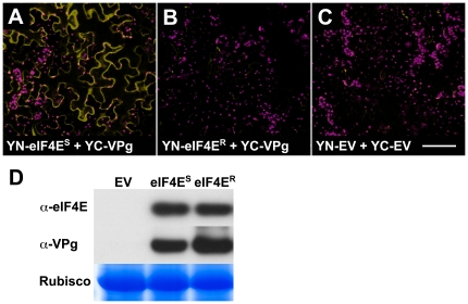
Figure 2. Pea eIF4ES interacts with PSbMV VPg in planta.
N. benthamiana leaves were co-infiltrated with constructs encoding TBSV P19 silencing suppressor [65] , PSbMV-P1 VPg fused to the C-terminal portion of YFP (YC-VPg) and either pea eIF4ES or eIF4ER fused to the N-terminal portion of YFP (YN-eIF4E). In control experiments, the equivalent empty expression vectors (YN-EV and YC-EV) were infiltrated in the presence of P19. YFP-specific fluorescence (yellow) and chloroplast autofluorescence (magenta) was recorded at 72 h post-infiltration by confocal microscopy. (A) A strong yellow fluorescence signal was detected in leaf epidermis following transient expression of YC-VPg and YN-eIF4ES (B) Expression of the YC-VPg+YN-eIF4ER combination resulted in a significantly lower yellow fluorescence signal. (C) Similar to that found for eIF4ER, yellow fluorescence was negligible following expression of the YN-EV+YC-EV vector controls. (D) Immunoblot analysis of total proteins confirmed that equivalent levels of eIF4ES, eIF4ER and VPg were present in infiltrated tissue. Size bar = 100 µm. In A–C data are representative of three independent experiments.

