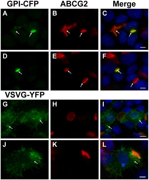
Figure 2. Apical localization of EVs in MCF-7/MR cells.
Cells were transiently transfected with GPI-CFP and VSVG-YFP constructs (green fluorescence), reacted with anti-ABCG2 antibody BXP-53 (red fluorescence) and DAPI and analyzed using Zeiss inverted Cell-Observer microscope at a magnification of ×630. Arrows denote the subcellular localization of GPI-CFP (A–F) and VSVG-YFP (G–L).

