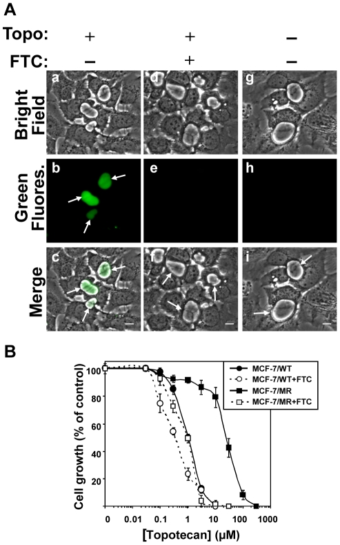
Figure 8. Inhibition of cell growth by topotecan and its accumulation in EVs.
(A) MCF-7/MR cells were grown in riboflavin-deficient medium for 7 days on dishes containing cover glass bottom. Then, cells were incubated with topotecan (5µM) for 24hr at 37°C in the presence (d–f) or absence (a–c) of FTC (10µM). Control cells were cultured in drug-free and riboflavin-deficient medium (g–i). Intravesicular accumulation of topotecan was analyzed as described in Materials and Methods. Arrows indicate the localization of EVs that lack or contain topotecan. (B) MCF-7 and MCF-7/MR cells were grown for 3 days, exposed to various concentrations of topotecan for 72h in the absence or presence of FTC (10µM), following which the cytotoxic effect was determined by the colorimetric XTT assay. Shown are the means of three independent experiments, each performed in triplicates ± SD. Topotecan IC50 values in MCF-7 and MCF-7/MR cells were 1.04±0.06 and 25.5±2.6, respectively.

