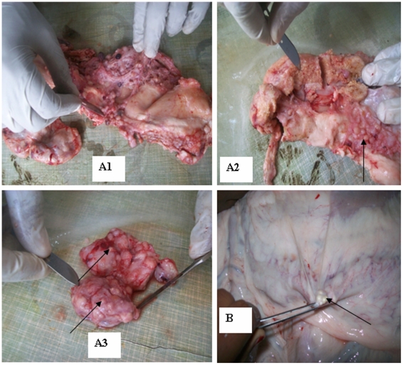
Figure 1. Tuberculous lesions from camels on different organs.
(A1) Disseminated and distinct tuberculous lesions in mediastinal parts of the lung. (A2) Tuberculous lesion in mediastinal lymph node and nodules on other parts as indicated by arrows. (A3) Tuberculous lesions in hepatic lymph node. The arrows show that pea-sized lesions throughout the lymph node. (B) Tuberculous lesion in mesenteric lymph nodes as indicated by arrow.

