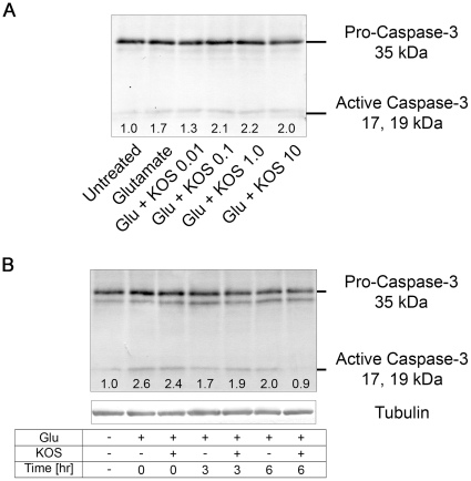
Figure 4. Effect of Koshu GSE on active caspase-3.
(A) Hippocampal neurons treated with 50 µM glutamate alone or in the presence of the indicated concentrations (in ng/ml) of KOS GSE for 30 min were analyzed for active-caspase-3 by Western blot. Numbers below the active caspase-3 bands indicate their relative intensities quantified by densitometric analysis. (B) Hippocampal neurons treated with 50 µM glutamate in the presence or absence of 1.0 ng/ml KOS GSE for 30 min and incubated in normal culture medium for 0, 3, or 6 hr were analyzed for active-caspase-3 by Western blot. Numbers below the active caspase-3 bands indicate their relative intensities quantified by densitometric analysis.

