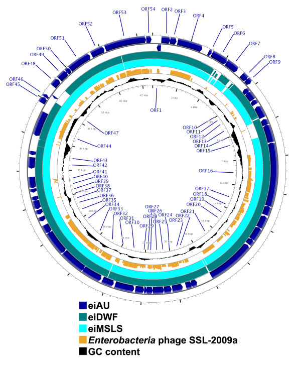
Figure 2.
Circular representation depicting the genomic organization of eiAU (two outermost circles, dark blue, showing each predicted ORF and its direction of transcription) and a tBLASTx comparison with the genomes of eiDWF (third circle from outside, green), eiMSLS (fourth circle from outside, light blue), and Enterobacteria phage SSL-2009a (fifth circle from outside, orange). The degree of sequence similarity to eiAU is proportional to the height of the bars in each frame. The %G+C content of eiAU is also depicted (sixth circle from outside, black). This map was created using the CGView server (Grant and Stothard, 2008).

