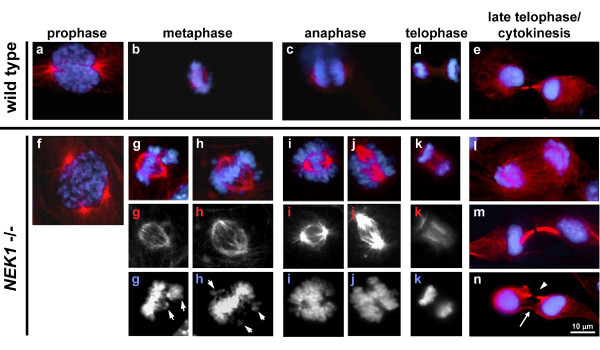
Figure 2.
NEK1 mutation results in disordered mitosis. Normal mitotic phases in primary passage 4 RTE cells from wild type littermate are shown in upper panels (a-e). DNA stained with DAPI fluoresces blue and secondarily recognized α-tubulin immunofluoresces red. In contrast, NEK1 -/- cells often undergo faulty and disordered mitosis (f-n), characterized by multipolar spindles (panel f), lagging chromosomes (panel g & h), improper directional movement of chromosomes or chromosome pieces during anaphase and telophase (panels i, j & k), and bizarre, incomplete types of cytokinesis (panel l, m & n). Grayscale panels are high-contrast images of the same panels above them, demonstrating individual staining for either α-tubulin or DAPI.

