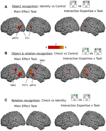
Figure 3. Neuroimagining data.
(A) The network of brain areas activated in recognition of chess object across all runs (whole session) – contrast Identity vs Control task (left side) and its interaction with expertise (right side). (B) The network of brain areas activated in recognition of chess object and their functions across all runs (whole session) – contrast Check vs Control task (left side) and its interaction with expertise (right side). (C) The network of brain areas activated in recognition of object functions across all runs (whole session) – contrast Check vs Identity task (left side) and its interaction with expertise (right side). The comparisons were based on p<.05 (corrected) and clusters of 5 or more voxels. The interaction between task and expertise in (A) and (C) were based on a lower threshold of p<.00001 (uncorrected). The significant areas included bilateral posterior middle temporal gyrus (pMTG), bilateral occipito-temporal junction (OTJ), right parieto-occipito-temporal junction (POTJ), and bilateral supramarginal gyrus (SMG). The MNI coordinates can be found below the labels of the ROIs in Figure 4.

