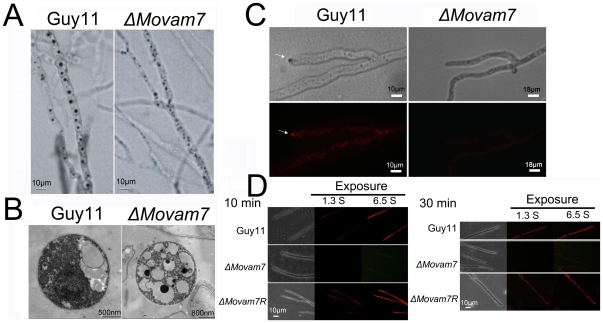
Figure 2. MoVam7 has a role in vacuole morphogenesis and the formation of the Spitzenkörper and endocytosis.
(A) Mycelia were stained with neutral red. At 14 hours, numerous small vacuoles were seen in the ΔMovam7 mutants (right panel), whereas large vacuoles of fewer numbers were present in the wild type strain. (B) Observation by transmission electron microscopy revealed numerous small, fragmented vacuoles in contrast to the large ones in the wild type hyphae. Bars represent 500 nm in the wild type strain or 800 nm in the ΔMovam7 mutant. (C) The wild type strain shows the presence of an intact Spitzenkörper (arrowheads) at the tips of the hyphae, which was missing in the ΔMovam7 mutants after exposure to FM4-64 staining for 10 min. Strains were grown for 2 days on CM-overlaid microscope slides before staining. (D) FM4-64 staining also revealed that the ΔMovam7 mutant was defective in endocytosis. Strains were grown for 2 days on the CM-overlaid microscope slides before adding FM4-64 and photographs were taken after 10 and 30 min exposure to FM4-64. Camera exposure is indicated in second.

