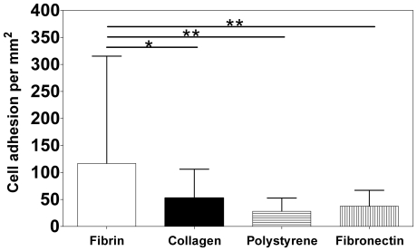
Figure 3. Adhesion of CD34+ cells to different substrates.
CD34+ cells were seeded on 24-well plates coated with fibrin gel, type I collagen gel (2.5 mg/mL) and fibronectin (50 µg/mL), and incubated for 3 h at 37°C. Polystyrene culture wells were used as control. After that time, the cells were washed and the attached ones were counted in six random microscope fields (×200 magnification) for each replicate. Results are average ± SD, n = 6. * and ** denote statistical significance (P<0.05 and P<0.01, respectively).

