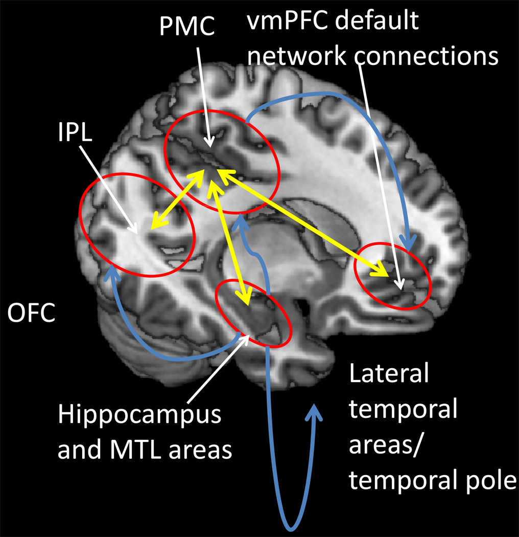
Figure 1.
Spread of the neuropathology in AD. Neurofibrillary tangles and neurodegeneration first appear in entorhinal cortex, and then in other medial temporal lobe (MTL) structures; fibrillary Aβ deposits and plaques first appear in transmodal areas [such as the posterior medial cortex (PMC), the inferior parietal lobule (IPL) and the lateral temporal lobe and temporal pole] that maintain reciprocal connections (illustrated by yellow arrows) with the entorhinal cortex. Spread of neurofibrillary tangles and neurodegeneration (illustrated by blue arrows) does not correlate with the spread of fibrillary Aβ deposition and plaque formation.

