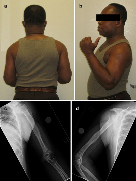
Fig. 5.
a A clinical photograph obtained at 4 months of follow-up after frame removal showing equal humeral lengths. b A clinical photograph obtained at 4 months follow-up after frame removal showing maximum elbow flexion and the actively extended thumb. c The AP radiographic view taken at 4 months follow-up after frame removal. d The lateral view obtained at 4 months follow-up after frame removal

