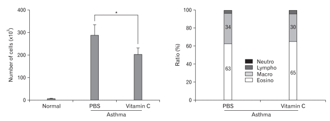
Fig. 2.
(left panel) Cell numbers in BALF. Mice were anesthesized and 1 ml of saline was infused into the lung through a needle in the trachea. The fluid was re-drawn after 1 min and centrifuged. Obtained cells were re-suspended, and cell count was done. Scanty cells were found in BALF from normal mice while many inflammatory cells were observed in BALF from asthma-induce mice. Vitamin C-treated mice showed less number of BALF cells than PBS-injected mice. A representative profile of three independent experiments. n=8 for each group. *P<0.05. (right panel) Differential cell counts of BALF cells from asthmatic mice. Cells were spread on a glass slide, stained with Wright's solution, and differentially counted under light microscope. In both experimental groups, eosinophils comprised about two thirds of total cells, and the other one third was almost macrophages. Lymphocytes and neutrophils were observerd with a very low frequency. These two groups showed no difference in cellular composition of BALF (P>0.05). Over 1,000 cells were counted in each group.

