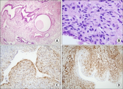
Fig. 3.
Microscopic finding. (A) The first biopsy of an endometrial polypoid lesion was composed of irregularly shaped glandular structures and loose fibromatous stroma (H&E, ×40). (B) The second biopsy of the endometrial mass was composed of glandular or cystic structures with stromal proliferation with a collar pattern and an intraluminal polypoid growth pattern (H&E, ×40). (C) Immunohistochemical (IHC) staining for CD10 shows positive stromal cells around epithelial structures (IHC, ×100). (D) Immunohistochemical staining for smooth muscle actin shows a positive reaction in most of the stromal cells (IHC, ×100).

