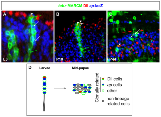
Fig. 4.
Cell migration pattern of clonally related cells. (A-C) Migration patterns of tub-MARCM clones (green) generated 72 hours AEL and analyzed in late L3 (A), P10 (B) and P44 (C). Dll-positive cells visualized with anti-Dll (red) and ap-positive cells with ap-lacZ (blue). White arrows in A-C mark ap-positive cells, green arrows mark GFP-positive cells negative for ap and arrowheads label Dll-positive cells. (D) Schematic representation of a larval `column' in the OPC. During pupation, these OPC-derived cells lose their columnar organization and become dispersed throughout the medulla cortex with non-lineage-related cells (gray cells).

