Fig. 4.
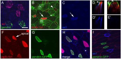
INC subtypes. (A) Analysis of Xenopus larval skin at stage 26 using two-label FISH probes for ae1 (red) and pendrin (green) identifies two approximately equal populations of INCs. (B-E′) Embryo injected at the 2-cell stage with mGFP RNA was fixed and stained at stage 26 with B1 (red) and ae1 (blue) antibodies. Note that INCs with strong apical B1 staining (B, arrow) also stain with ae1 (C, arrow) whereas those with basolateral B1 staining (B, arrowhead) do not. z-section through cells with apical B1 staining (compare arrowheads in D and E) shows that ae1 localizes basolaterally (D,D′), whereas in cells with cytosolic or basolateral B1 staining ae1 is absent (E,E′). (F-H) pendrin-GFPtr embryos were fixed at stage 26 and stained with antibodies to B1 (red) and E-cadherin (blue). Shown is B1 staining (F), GFP expression (G), or a merge of all three channels (H). (I) pendrin-GFPtr embryos were injected with mRFP RNA (red) to label cell boundaries and stained with ae1 antibody (blue). CC, ciliated cell. Asterisk marks a small population of INCs that do not stain with or express ae1, pendrin or B1. Scale bar: 20 μm.

