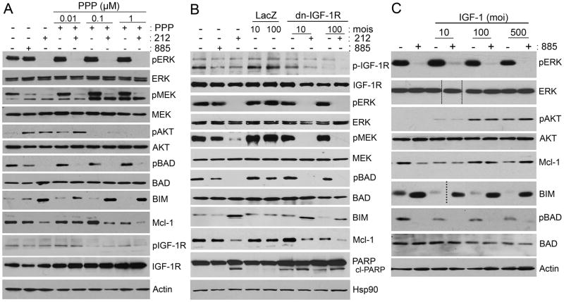
Figure 6. IGF-R mediates PI3K signaling in BRAF-inhibitor resistant cells.
(A) Mel1617-R cells were treated with increasing concentrations of PPP (μM) as single agent or in combination with 0.1 μM 212. The effect of IGF-1R inhibition on MAPK, AKT, and Bcl-2 family proteins was assessed by immunoblotting. (B) Mel1617-R cells were infected with adenoviruses encoding dominant negative (dn) IGF-1R at 10 or 100 MOI, or LacZ as a negative control. Infected cells were treated 48h post infection with 0.1 μM 212 or left untreated. Cells lysates were analyzed by immunoblotting; cl-PARP, cleaved PARP. (C) Parental Mel1617 cells were infected in serum-free medium with adenoviruses encoding IGF-1 at 10, 100 or 500 MOI. Infected cells were serum starved for 48 h and then treated with 1 μM 885 or left untreated. Cells lysates were analyzed by immunoblotting. Doted lines indicate where blot was cut to remove an empty lane. See also Figure S5.

