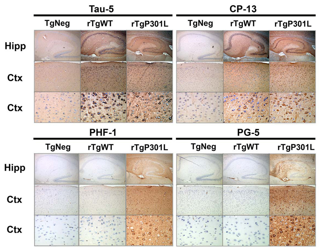
Figure 7. Pathological tau species do not accumulate in rTgWT mice.
At 5 months of age (approximately when memory impairments begin, but before neuron loss), immunohistochemical studies revealed that cortical and hippocampal neurons in rTgWT mice were rarely immunoreactive for hyperphosphorylated tau species associated with disease. Detection of total tau (human and mouse) with the Tau-5 antibody in hippocampal and cortical neurons of rTgWT and rTgP301L mice. At 5 months, an immunoreactive signal for tau hyperphosphorylated at residue S202 (CP-13) was observed in the cell bodies of a small number of rTgWT hippocampal and cortical neurons. In contrast, CP-13 labeled-cell bodies and neurites were observed in a majority of rTgP301L hippocampal and cortical neurons. Strikingly, positive immunoreactive labeling for tau hyperphosphorylated at S409 (PG-5) and S396/S404 (PHF-1) was observed in hippocampal and cortical cell bodies and neurites of 5 month-old rTgP301L, but not rTgWT, mice. Nuclei were stained with DAPI. For each antibody panel, magnification was x5 (top row), x10 (middle row) and x40 (bottom row). Hipp, hippocampus; Ctx, cortex.

