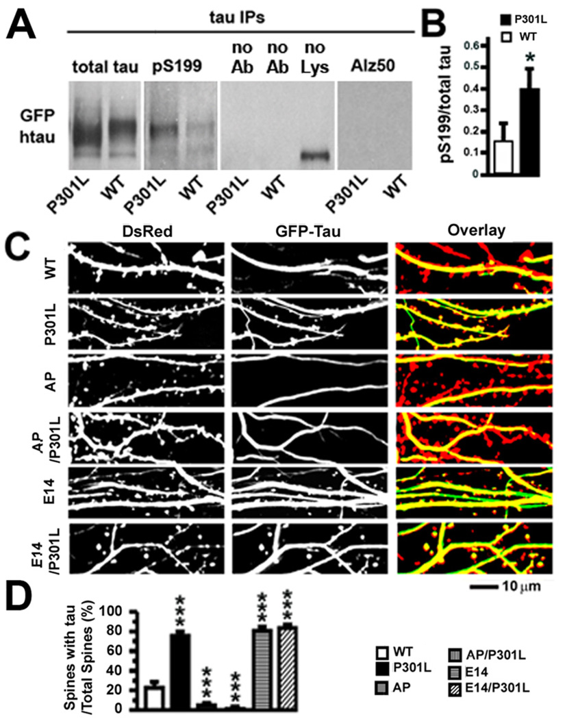
Figure 8. Hyperphosphorylation of P301L htau and effects of proline-directed phosphorylation on tau accumulation in dendritic spines of transfected rat hippocampal neurons.
(A) Levels of phosphorylated htau in rat neurons expressing WT or P301L htau. Expression of total htau and phosphorylated S199 (pS199) htau proteins after immunoprecipitation of total htau with the Tau-13 antibody from 21–28 DIV primary rat hippocampal neurons expressing WT or P301L htau. Changes in tau conformation and phosphorylation states associated with late tau pathology were also examined with the Alz-50 antibody. In the middle panel, omission of the Tau-13 antibody (no Ab) or tissue lysate (no Lys) revealed no specific immunoblot signal for total htau. (B) There was more pS199, normalized to total tau, in the P301L htau-expressing neurons. n = 3 sample preparations. (C) Images of living rat neurons co-expressing DsRed and six different GFP-tagged htau proteins (WT, P301L, AP, AP/P301L, E14 or E14/P301L). In neurons expressing WT, AP and AP/301L htau most DsRed-labeled spines were devoid of htau. In neurons expressing P301L, E14 and E14/P301L htau most spines contained htau. (D) Quantification of total spines and htau-containing spines in neurons co-expressing DsRed and GFP-tagged htau. A higher percentage of spines contained htau in neurons expressing P301L, E14 and E14/P301L htau (compared to WT htau), and a lower percentage of spines contained htau in neurons expressing AP and AP/P301L (compared to WT htau). n = 25–28 neurons per group. t-test (B), ANOVA followed by Bonferroni post-hoc analysis (D), *p < 0.05, ***p < 0.001.

