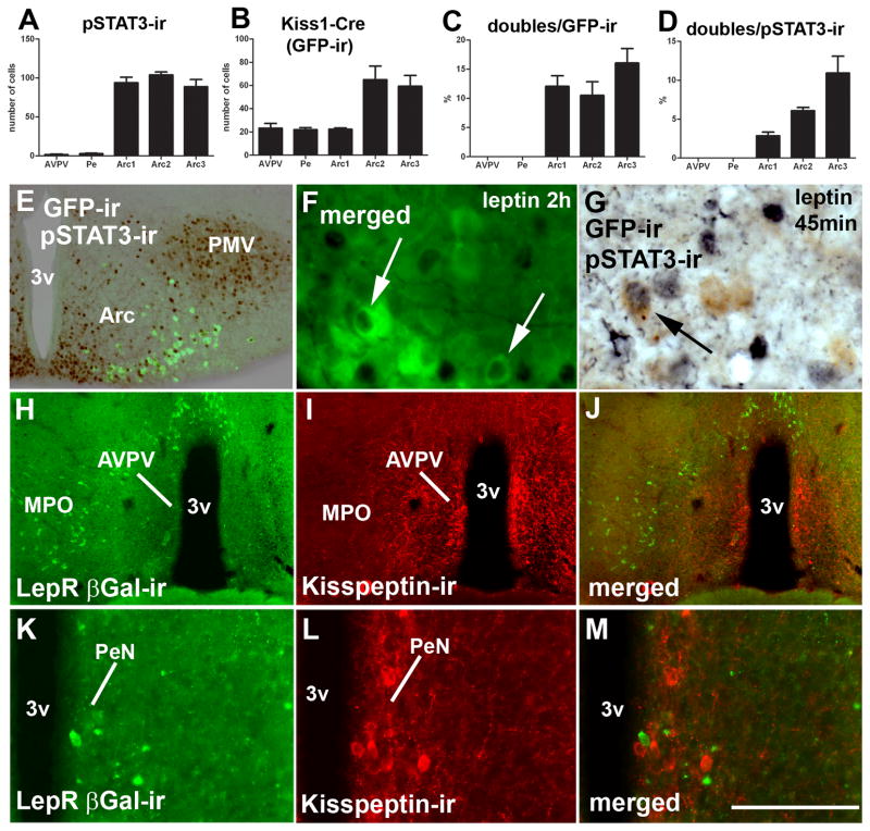
Figure 7.
Distribution of Kiss1 neurons responsive to leptin. A, bar graph showing number of leptin-induced phosphorylation of STAT3 immunoreactivity (pSTAT3-ir) in hypothalamic nuclei which express Kiss1 reporter gene (anteroventral periventricular nucleus/AVPV, anterior periventricular nucleus/PeN, 3 rostro-to-caudal levels of the arcuate nucleus Arc1-3); B, bar graph showing the number of neurons expressing Kiss1 reporter gene (green fluorescent protein immunoreactivity/GFP-ir); C, percentage of Kiss1 neurons expressing pSTAT3-ir; D, percentage of pSTAT3-ir neurons expressing Kiss1 reporter gene. Females on diestrus from line J2-4 were used in this experiment. E, fluorescent and brightfield photomicrographs showing distribution of leptin-induced pSTAT3-ir and Kiss1 neurons (reporter gene), in a caudal level of the Arc. F–G, fluorescent and brightfield photomicrographs showing Kiss1 neurons (reporter gene) expressing leptin-induced pSTAT3-ir (black nucleus, arrows). H–M, fluorescent photomicrographs showing the distribution of leptin receptor reporter gene (LepR, βGalactosidase immunoreactivity/βGal-ir) in the preoptic area. Note the reduced number of neurons expressing LepR reporter gene in the AVPV (H–J) and PeN (K–M), and the absence of colocalization between LepR and kisspeptin immunoreactivity. Abbreviations: 3v, third ventricle; MPO, medial preoptic nucleus; ox, optic chiasm; PMV, ventral premammillary nucleus. Scale bar: E–F = 200 μm; G = 50 μm; H–J, 400 μm; K–M, 100 μm.

