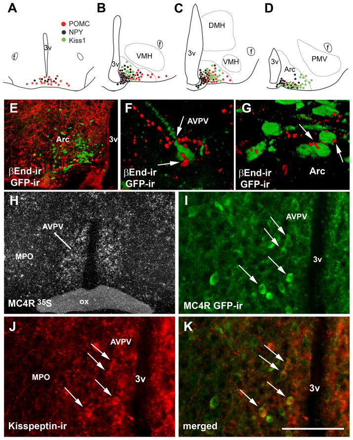
Figure 9.
Topographic distribution of Kiss1 neurons and their innervation by melanocortins. A–D, schematic drawings showing the topographic distribution of Kiss1 neurons (green dots, line J2-4), proopiomelanocortin neurons (red dots) and neuropeptide Y neurons (black dots) in the retrochiasmatic area (RCA) and 3 rostro-to-caudal levels of the arcuate nucleus (Arc). E, fluorescent photomicrograph showing the segregated distribution of neurons expressing Kiss1 reporter gene (green fluorescent protein immunoreactivity/GFP-ir, green cytoplasm) and βEndorphin immunoreactivity (βEnd-ir, a POMC product, red cytoplasm). F–G, fluorescent photomicrographs showing close appositions between βEnd immunoreactive fibers and GFP immunoreactive neurons, in the anteroventral periventricular nucleus (AVPV, F) and in the Arc (G). H, darkfield photomicrograph showing expression of melanocortin 4 receptor (MC4R) mRNA (hybridization signal, silver grains) in the AVPV of a female mouse. I–K, fluorescent photomicrographs showing the colocalization of MC4R reporter gene (GFP-ir) and kisspeptin immunoreactivity (kisspeptin-ir) in the AVPV (arrows). Abbreviations: 3v, third ventricle; MPO medial preoptic nucleus; ox, optical chiasm. Scale bar: A–D = 600 μm; E, H = 400 μm; I–K = 100 μm, F–G, 50 μm.

