
Fig. 4.
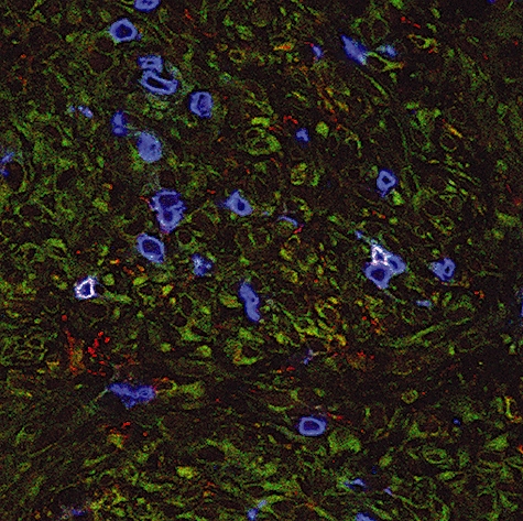
In situ detection of natural killer (NK) T cells in tumour from patient B2. Cryosections of tumour tissue were stained with anti-T cell receptor (TCR) Vα24-fluorescein isothiocyanate (green) in combination with anti-TCR Vβ11-phycoerythrin (red) and anti-CD3 (blue), as described in Materials and methods. Triple-positive NK T cells are detected by a white membrane staining. Renal tumour cells show some aspecific background staining in green. Original magnification × 400.