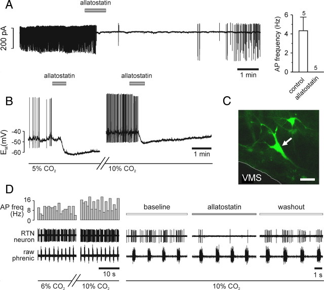
Figure 2.
Acute silencing of individual ventrolateral brainstem Phox2b neurons expressing AlstR-EGFP by allatostatin. A, Representative cell-attached recording from an EGFP-positive ventrolateral brainstem neuron (left). As the mean data (right) show, such cells spontaneously fire action potentials under normocapnic conditions. Bath-application of 1 μm allatostatin rapidly and reversibly abolished action-potential firing in all cells tested. B, Representative whole-cell current-clamp recording from an EGFP-positive neuron. Response to 1 μm allatostatin at normocapnia (left), followed by a washout of ∼30 min for recovery and change of solution by the response to allatostatin in 10% CO2 (right). In both conditions, allatostatin rapidly hyperpolarized the cell, leading to cessation of action potential firing. C, Cell recorded in B (confocal z-stacked image). Scale bar, 40 μm. D, Representative extracellular recording of CO2-sensitive RTN neuron in the in situ brainstem–spinal cord preparation. Chemosensitivity of the neuron is shown by the increased firing frequency at 10% CO2. Allatostatin application produced a significant decrease in spiking activity of RTN neurons at high CO2 (n = 4). AP freq, Action-potential frequency.

