
Figure 3. Systemic MB reduced the metabolic lesion in the PCC produced by AZ.
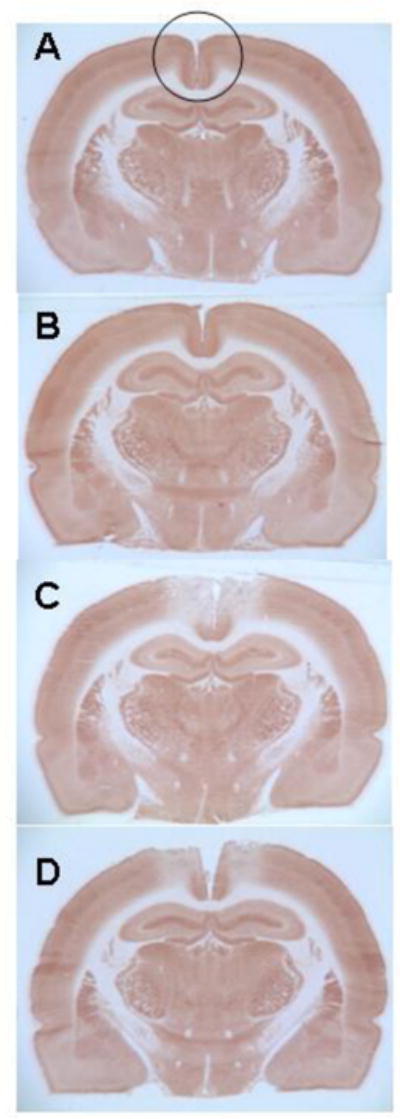
Representative photomicrographs of comparable cytochrome oxidase histochemistry-stained sections at the level of the AZ injection sites in the PCC to illustrate primary histochemical data. The photomicrographs show entire sections to illustrate that the inhibition of cytochrome oxidase activity produced by AZ injections was limited to the PCC (area inside the circle) and did not involve other regions. Sections from each group were stained in parallel and the darkness (optical density) of the stained tissue was directly proportional to its cytochrome oxidase activity, as calibrated in each staining batch with tissue standards of known enzymatic activity measured biochemically (for histochemistry details refer to Gonzalez-Lima and Cada, 1998). A) Control brain. Section from a sham operated rat not receiving any injections into the PCC, illustrating normal cytochrome oxidase histochemistry. B) Control vehicle-injected brain. Section from a sham operated rat receiving bilateral vehicle injections into the PCC, illustrating minimal effects on the cytochrome oxidase histochemistry. C) AZ injected brain. Section from an operated rat that received bilateral AZ injections into the PCC, illustrating reduced cytochrome oxidase histochemical activity in the PCC. D) AZ+MB injected brain. Section from an operated rat that received bilateral AZ injections into the PCC plus systemic MB, illustrating attenuated AZ effects on the cytochrome oxidase histochemical activity of the PCC.