
Figure 5. Systemic MB did not prevent the reduction in neuronal density produced by AZ in the dorsal PCC.
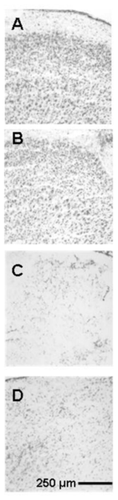
Representative photomicrographs of high magnification Nissl-stained sections used to count cells at the AZ injection sites in the dorsal PCC to illustrate primary cell counting data. The maximal AZ effect was in the dorsal PCC targeted by the injections, and systemic MB did not prevent neuronal loss in this region. A) Control brain. Section from a sham operated rat not receiving any injections into the PCC, illustrating normal cell density. B) Control vehicle-injected brain. Section from a sham operated rat receiving bilateral vehicle injections into the PCC, illustrating minimal effects on the cell density. C) AZ injected brain. Section from an operated rat that received bilateral AZ injections into the PCC, illustrating discoloration and neuronal loss in the dorsal PCC. D) AZ+MB injected brain. Section from an operated rat that received bilateral AZ injections into the PCC plus systemic MB, illustrating AZ effects on neuronal density in the PCC that were not statistically different from the AZ group.