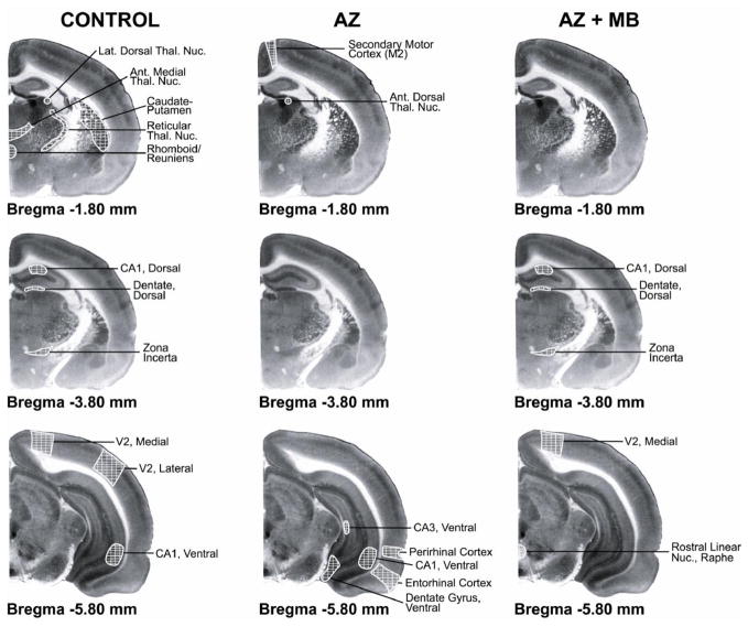
Figure 6. Systemic MB partially prevented the disruption in PCC functional network induced by AZ.
Coronal brain sections stained with the cytochrome oxidase histochemistry technique depict the number and anatomical distribution of brain regions showing functional networks with the posterior cingulate cortex (PCC) (white cross-hatching). In control conditions, the metabolic capacity of the PCC was significantly correlated with that of thalamic, hippocampal and extrastriate regions. Except for the ventral CA1, AZ induced a complete loss of this pattern of PCC functional networks, and strengthened PCC connectivity with a different set of hippocampal and parahippocampal regions, as well as with the secondary motor cortex. In contrast, MB not only prevented this new pattern of PCC connectivity, but also partially preserved the PCC connectivity with the hippocampus and the extrastriate cortex as seen in the control. MB also induced coupling between the PCC and the raphe nuclei.

