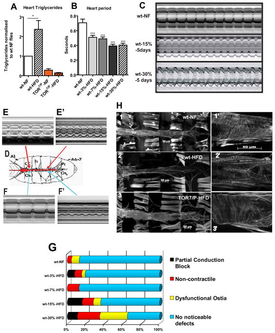
Figure 2. HFD treatment causes cardiac dysfunction.
A) Triglyceride content from female hearts on NF and HFD for 5 days (normalized to wt NF hearts). TOR7/P mutant flies were examined under the same food conditions. At least 3 independent experiments were done for each time point for all TG experiments (with approximately 90 hearts were used for each condition and genotype). wt type flies showed an increase in TGs (* = p<0.05) while the TOR mutants had significantly lower levels of TGs that did not increase on a HFD. B) Bar graph representation of changes in heart period for a population of approximately 24 flies hearts from each food type. A dose dependent effect is shown for the increase in dietary fat content (***p<0.0001). A minimum of 22 flies were used for each dietary condition. C) M-mode traces prepared from high speed movies of semi-intact preparations on NF, 15%, and 30% HFDs for 5 days. Note the changes in the M-mode traces as the concentration of fat increases. Red bars indicate systolic and diastolic diameters. D) A sketch of a dissected semi-intact heart preparation. Arrows indicate to the area the corresponding M-modes shown in E–F′. E–E′) Representation of partial conduction blocks of hearts using M-modes from different portions of the same heart. E) M-mode from the anterior portion of the heart displaying a regular heart beat. E′) M-mode from the posterior portion of the same heart displaying a faster and erratic heart beating pattern, compared to I. F–F′) Representation of a portion of the heart that is non-contractile hearts using M-modes of different heart regions. F) M-mode taken from the anterior portion of the heart displays regular beating pattern. F′) M-mode taken from the posterior portion of the same heart showing poor or no contractions. G) Side bar graph of heart phenotypes seen under HFD conditions with increasing amount of fat. The heart phenotypes used in this graph are partial conduction blocks, non-contractile myocardial cells, dysfunctional ostia and no noticeable defects. The instances of all 3 phenotypes increase in a fat dose-dependent fashion. A minimum of 26 heart movies were analysed for each dietary condition. H) Fluorescent micrographs of hearts on NF and HFD for 5 days at 10x and 25x optical magnification. Adult hearts are stained with AlexaFlourR 594 phalloidin. 1) Adult heart of a wt flies on NF. 1′) Magnified anterior NF-fed wt heart region. Note the regular arrangements of myofibrillar organization. 2) Adult heart of a wt flies on HFD for 5 days. Note the degeneration of the regular myofibrillar structure and the decrease in heart tube diameter. 2′) Magnified anterior wt heart region on HFD. Note the disorganization of the circular myocardial myofibrils. 3) Adult heart of a TOR7/P mutant on a HFD for 5 days. Note that there is little to no degradation of heart structure and the diameter is not constricted. 3′) Magnified anterior heart region of a TOR7/P mutant. There is little change in myocardial structure under HFD conditions compared to NF-fed wt (1′).

