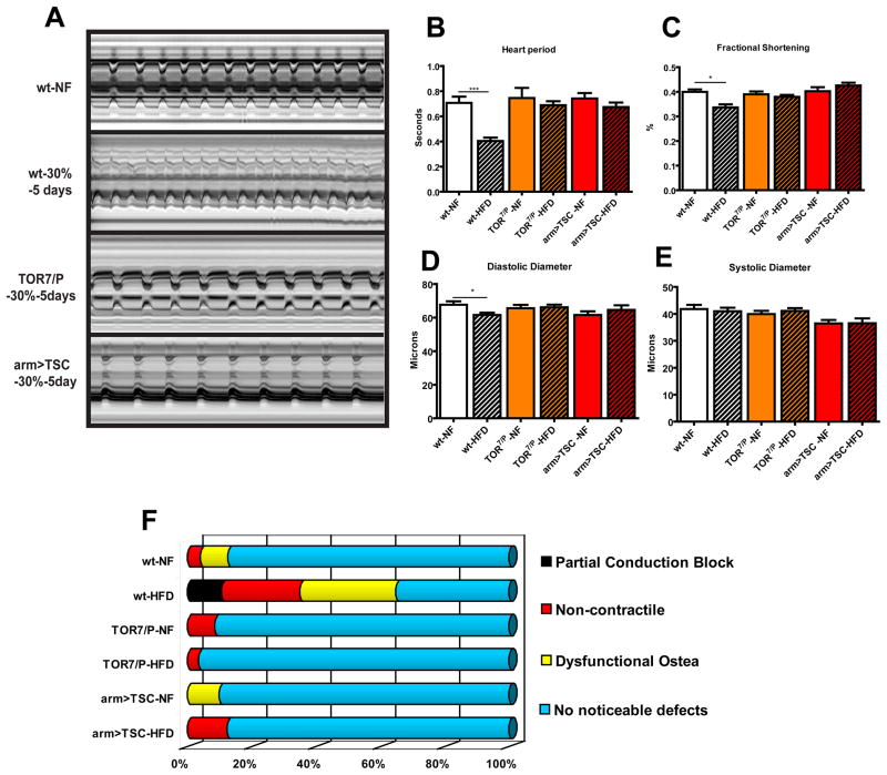
Figure 4. Reducing TOR function prevents HFD-induced obesity cardiac dysfunction.
A) M-mode traces of dissected wt flies on NF and 30% HFD, TOR7/P mutants and arm>TSC1-2 on NF and 30% HFD. No significant change was seen in TOR mutant nor arm<TSC heart M-modes on HFD when compared to wt on NF. B) Bar graph of cumulative heart periods for wt, TOR mutants (n=25, 36 respectively) and arm>TSC1-2 flies (n= 20 (NF), 21 (HFD)) under NF and HFD conditions. No change in heart period from wt under NF was seen in TOR mutants and arm>TSC1-2 flies under HFD conditions. C) Bar graph of changes in Fractional Shortening. A decrease in Fractional Shortening in wt flies was seen after 5 days on a 30% HFD (p<0.001). While no change was seen in fractional shortening in neither the TOR mutants nor the arm>TSC1-2 flies under a HFD. D) Bar graph of combined diastolic diameter data of wt and TOR mutant and arm>TSC1-2 hearts under NF and 30% HFD conditions. No change was seen in Diastolic Diameter in TOR mutants and arm>TSC1-2 flies under a HFD. E) Bar graph of systolic diameter of fly heart as in (F). No decrease in Systolic Diameter was seen after 5 days on a 30% HFD for any of the strains tested. F) Graphical representation of heart phenotypes as in Fig. 2G. No significant change the heart phenotype of TOR7/P mutants or with systemic TSC1-2 overexpression under HFD could be detected, when compared to wt flies. A minimum of 20 individual fly heart movies were analyses.

