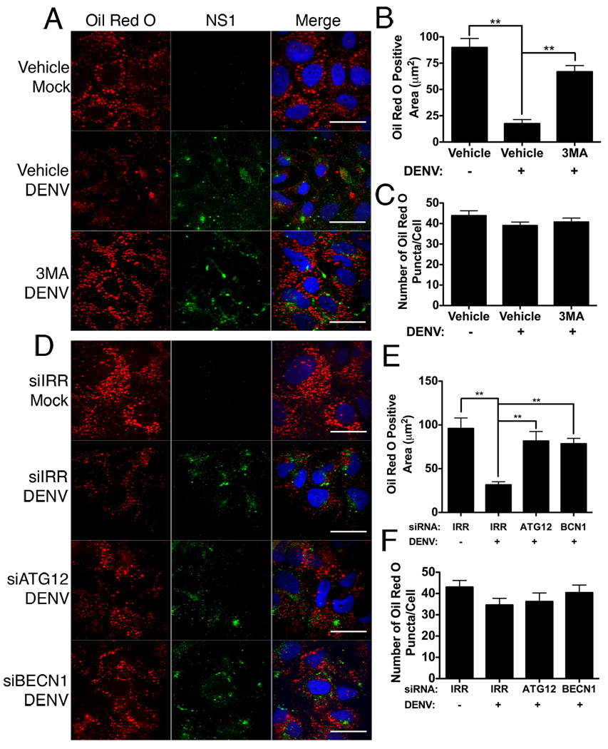
Figure 3. Inhibition of autophagy prevents depletion of lipid droplets in DENV-infected cells.
(A) Huh-7.5 cells were mock- or DENV-infected at an MOI of 2 for two hours, then treated with 2.5mM 3MA or vehicle control. Cells were fixed 48HPI and stained with Oil Red O to visualize lipid droplets and an antibody against DENV NS1 (green). (C) Alternatively, Huh-7.5 cells were treated with siRNAs targeting an irrelevant HCV sequence (IRR), ATG12, or beclin1 (BECN1) for 24 hours, and then infected with DENV for 48 hours at an MOI of 2. Respective Image J quantification of total area staining positive for oil red O (B,E) and the total number of oil red O puncta per cell (C,F). Values represent the average of at least eight cells per treatment. **p≤0.001. Scale bar = 30µm.

