
Fig. 2.
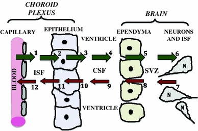
Central role of CP–CSF in exchanging materials with brain: ions, water, and organic molecules filter passively out of choroidal capillaries (arrow #1) into interstitial fluid (ISF). This is the first step in the material flow by distributional nexus to the brain (green arrows). Solutes diffuse through ISF up to basolateral membrane of epithelium. Active mechanisms in membranes transfer solutes sequentially across the basolateral (#2) and apical membranes (#3), into ventricular CSF. As CSF flows (#4) from ventricles to cisterna magna, a small fraction of the CSF-borne substances diffuse across ependyma (#5) into periventricular brain or are taken up by specialized ependymal cells. Ependyma-penetrating substances diffuse through brain ISF (#6) for transport into neurons (N); BBB, not depicted, is interspersed among neurons. Material inflow to the CNS interior thus sequentially involves CP, CSF, ependyma and brain. In the opposite direction, there is a reverse nexus (red arrows) for catabolites or injury products (as in TBI) released by neurons/glia into brain ISF. Accordingly, cerebral catabolites such as homovanillic acid diffuse through ISF (arrow #7), and down a transependymal concentration gradient into CSF (#8). By bulk flow of CSF (#9), catabolites are convected to subarachnoid space (not depicted) or to CP for active removal from the ventricles (#10), and then extrusion transport across basolateral membrane (#11). Therefore, some endogenous solutes or injury products end up being cleared passively into blood of microvessels (#12) and venules draining CP. Overall CSF is simultaneously a source (#1 to #6) and a sink (#7 to #12) for distributing molecules, depending on prevailing concentration gradients between ventricular CSF and brain ISF. As such, the CSF and bordering cells constitute a nexus for mediating trophic (CSF to brain) and excretory (brain to CSF) fluxes