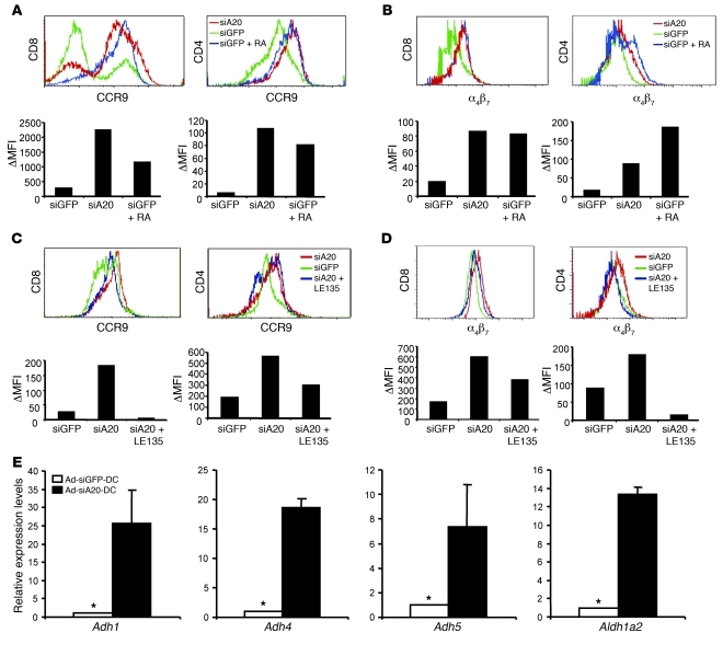
Figure 6. Ad-siA20-BM-DCs induced gut-homing receptors on T cells in vitro.
(A and B) Surface expression of CCR9 (A) and α4β7 (B) on CD8+ OT-I or CD4+ OT-II T cells after in vitro coculture with Ad-siA20- or Ad-siGFP-BM-DCs pulsed with H-2Kb/OT-I or OT-II peptide (10 μg/ml) (5 × 10⁴ cells/well) in the presence or absence of RA (Sigma-Aldrich, 100 nM); data are from 1 of 2 repeated experiments. (C and D) Reduced expression of CCR9 (C) and α4β7 (D) by blocking RA receptors. Surface expression of CCR9 and α4β7 on CD8+ OT-I or CD4+ OT-II T cells was examined by flow cytometry after coculture with Ad-siA20- or Ad-siGFP-BM-DCs pulsed with H-2Kb/OT-I or OT-II peptide in the presence or absence of the RA receptor antagonist LE135 (1 μM, Tocris Bioscience). Data from 1 of 2 repeated experiments are presented. (E) Increased expression of RA-producing enzymes by Ad-siA20-BM-DCs. qRT-PCR was used to analyze Ad-siA20-BM-DCs and Ad-siGFP-BM-DCs after stimulation with LPS for 12 hours. The relative mRNA levels of the RA-producing enzymes from 1 of 2 repeated experiments are presented. *P < 0.05, Ad-siGFP-BM-DCs versus Ad-siA20-BM-DCs.

