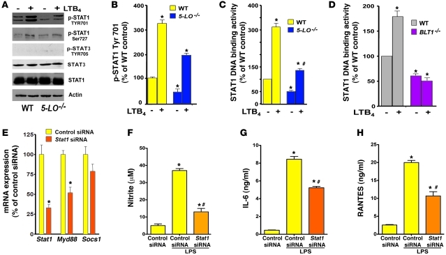
Figure 4. LTB4/BLT1-induced STAT1 activation is required for MyD88 expression and TLR4 responses.
(A) WT and 5-LO–/– macrophages were treated for 30 minutes with or without LTB4, and pSTAT1 (Tyr701), pSTAT1 (Ser727), total STAT1, pSTAT3 (Tyr705), total STAT3, and actin were determined by immunoblotting. (B) Densitometric analysis of pSTAT1 (Tyr701) protein levels in 5-LO–/– macrophages, normalized for total STAT1 and expressed as percent of WT control cells. (C and D) WT and 5-LO–/– macrophages (C) and WT and BLT1–/– cells (D) were incubated for 30 minutes with or without LTB4, and nuclear STAT1 activity was determined as described in Methods. (E) WT macrophages were treated for 48 hours in the presence of scrambled siRNA (control siRNA) and Stat1 siRNA, and the expression of Stat1, Myd88, and Socs1 mRNA was determined by real-time RT-PCR and expressed relative to control siRNA values. (F) NO, (G) IL-6, and (H) RANTES secretion in Stat1-silenced WT macrophages incubated with or without LPS for 24 hours. Data represent mean ± SEM from 3 individual experiments, each performed in triplicate. (B–D) *P < 0.05 versus unstimulated WT; #P < 0.05 versus stimulated WT. (E–H) *P < 0.05 versus control siRNA; #P < 0.05 versus LPS-challenged control siRNA.

