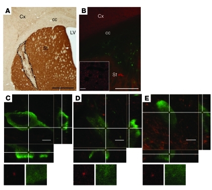
Figure 6. In vivo transmission of α-syn from mouse brain to a graft of dopaminergic neurons.
(A) Representative coronal section from grafted human α-syn–overexpressing mouse stained with an antibody against TH. The dashed line delineates the transplant of TH-positive neurons in the host striatum. (B) Grafted neurons are identified by TH staining (green) within the human α-syn–positive (red) striatum of the host. The inset shows high magnification of human α-syn–positive accumulations in the host striatum. (C–E) Confocal 3D reconstruction of wild-type TH-positive cells (green) in transgenic mice overexpressing human α-syn. The cross points on human α-syn–positive dots (red) present within the transplanted cells. Reconstructed orthogonal projections are presented as viewed in the x-z (bottom) and y-z (right) planes. Cx, cortex; cc, corpus callosum; LV, lateral ventricle; St, striatum. Scale bars: 1,000 μm (A); 500 μm (B); 10 μm (B, inset); 5 μm (C–E). Original magnification, ×1915 (C–E).

