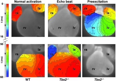
Figure 3. Typical example of an activation pattern in a wild-type and Tbx2–/– hearts at E14.5.
In the wild-type heart, activation starts in the atria, and after a delay of 50 ms the ventricles are activated within 3 ms after the first moment of activation of the apex of the left ventricle. In the middle panel, an activation pattern of a Tbx2–/– heart is shown. The activation starts in the atria, and after a normal AV delay, the ventricles are activated from apex to base, after which the atria are activated for the second time via the left side. The right panel shows an example of ventricular preexcitation in a Tbx2–/– heart. The activation starts in the atria, after which the base of the left ventricle is activated with an AV delay of 8 ms. Complete activation of both the left and right ventricle is within 15 ms. Original magnification, ×5.

