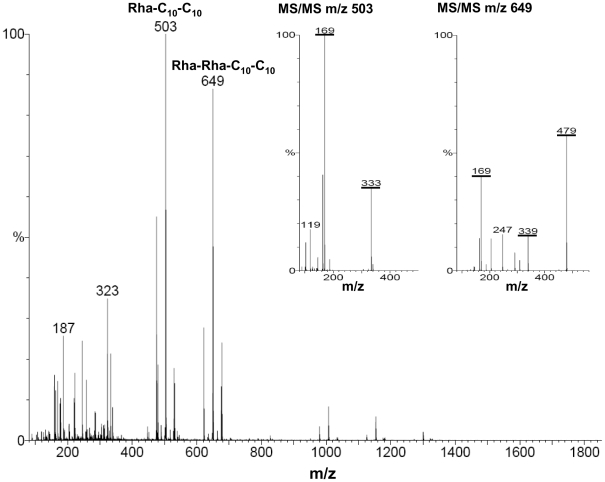
Figure 4. Electrospray-ionization-mass-spectrum of rhamnolipids isolated from the clinical isolate PA264.
Analyses of the rhamnolipid preparation were performed in the negative ionization mode. The ion at m/z 503 corresponds to the mono-rhamnolipid Rha-C10-C10 as revealed by MS/MS analyses of m/z 503 displayed in the inset. The ion at m/z 649 corresponds to the dirhamnolipid Rha-Rha-C10-C10 as revealed by MS/MS analyses of m/z 649 displayed in the inset. The corresponding and characteristic ions formed by collision-induced fragmentation are underlined.

