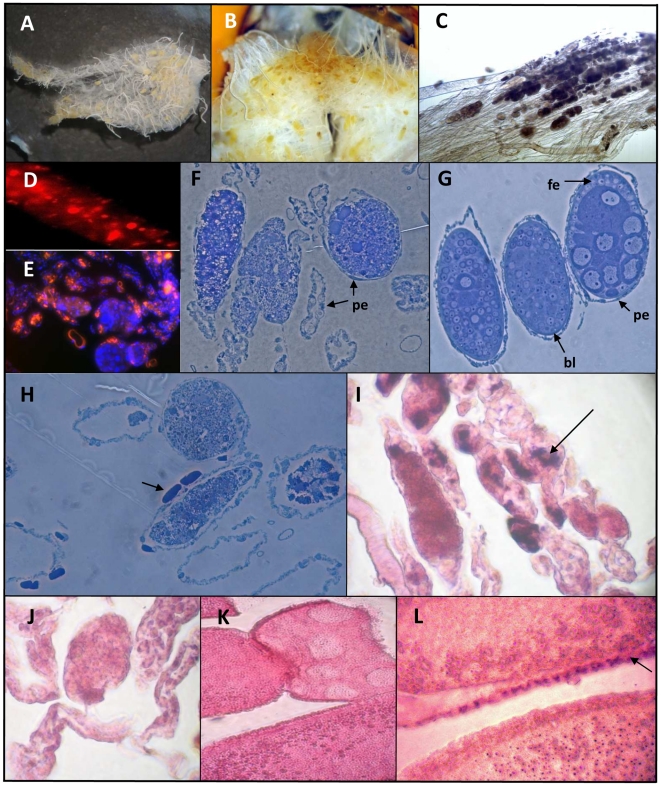
Figure 1. Histological analysis of the queen ovary.
A and B: Stereomicroscopic observation of ovaries displaying yellowish colorations in the ovarioles. C: Clumps in the terminal filament of ovarioles observed by phase contrast microscopy (Olympus MVX 10). D: Propidium iodide staining of an ovariole showing nuclear condensation in the terminal filament (×600). E: general view of the apex of an ovary double stained with propidium iodide (red) and DAPI (blue). F, G and H: Toluidin blue staining of 1 µm thick cuttings from the germinal region of two ovaries. F and H: ovarioles displaying degeneration process showing detail of the disorganization of follicles surrounded by several empty ovarioles; H: presence of crystalline arrays in the peritoneal epithelium (arrow). G, normal ovary: detail of three follicles at different developmental stages. The nurse cells chamber and the oocyte chamber differentiation appear on the follicle located on the right. fe: follicular epithelium; pe: periteoneal epithelium; bl: basal lamina. I–L: In situ hybridization assays performed from paraffin embedded tissues for the detection of DWV/VDV-1 RNA. I and J: staining performed from the terminal filament region of an ovary using either antisense (I) or sense (J) probe. K and L: in situ analysis performed from the vitellarium showing DWV/VDV-1 RNA staining in the periphery of an oocyte (Arrow) with the antisens probe (L); K: control sense probe. Slices were counterstained with eosin Y.

