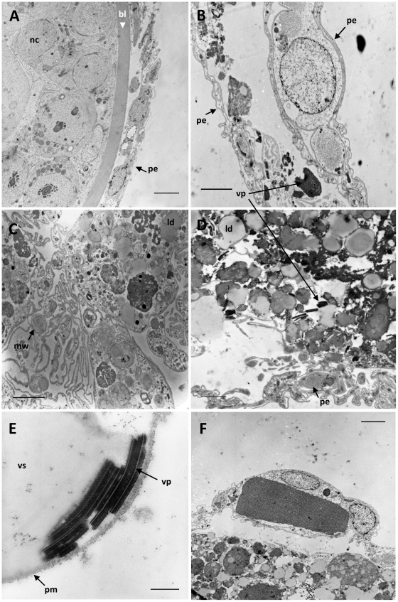
Figure 2. Ultrastructural observations in the germarium.
A: TEM analysis of a normal follicle showing nurse cells (nc), the thick basal lamina (bl) and the peritoneal epithelium (pe). B: TEM analysis of an empty ovariole with presence of viral particles (vp) and cellular debris lined by the peritoneal enveloppe (pe). C and D: TEM analysis showing cellular degeneration of follicular cells with myelin whorls (mw), lipid droplets (ld), viral particles (vp) and peritoneal envelope (pe). E: TEM observation of viral particles (vp) and the virogenic stroma (vs) near the plasma membrane (pm) of a follicular cell. F: detail of a crystalline matrix in a peritoneal epithelial cell. Bars in electron micrographs represent 5 µm, 3 µm, 2 µm, 2 µm, 250 nm and 3 µm in figures A, B, C, D, E and F, respectively.

