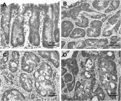
Fig. 5.
Immunohistochemical analysis of β-catenin in colonic tissue. hCYP1A transgenic mice were treated with 200 mg/kg PhIP and 1.5% DSS and sacrificed 10 weeks after PhIP administration. (A) Normal colon mucosa with brown staining in the membrane and cytoplasm, (B) high-grade dysplasia with brown staining of β-catenin in the nuclei, (C) nuclear staining of β-catenin in colonic adenomas and (D) intense nuclear staining for β-catenin in colonic adenocarcinomas.

