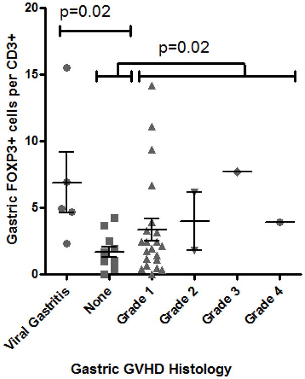
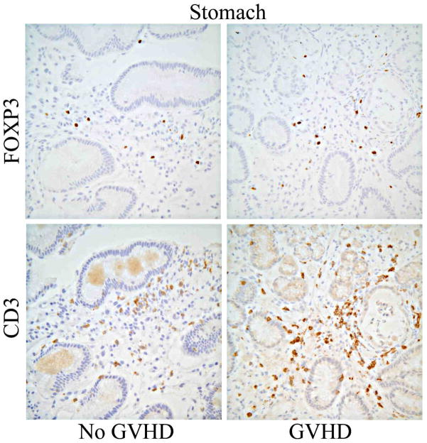
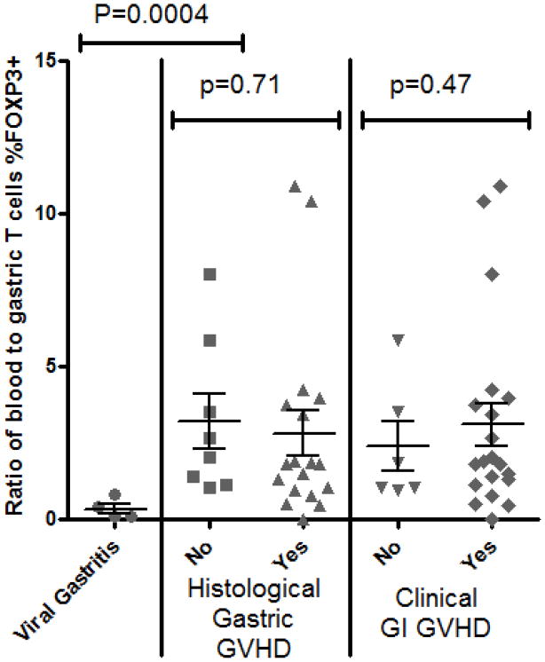
Figure 3. Gastric mucosal Tregs are not decreased in GVHD.

A: Representative histology of gastric biopsies from patients with (right panels) versus without (left panels) gastric GVHD, stained immunohistochemically for FOXP3 (upper panels) or CD3 (lower panels). B: The FOXP3+ to CD3+ ratio (expressed as a percent) of serial sections of gastric biopsies was compared based upon the histological grade of GVHD severity within those biopsies. P-value shown is for comparison between pooled specimens with grade I–IV GVHD, versus specimens with no GVHD (“none”) or viral gastritis. C: The percent of T cells expressing FOXP3 (per CD3+ cells in gastric biopsies, and per pooled CD4+ and CD8+ in blood) was expressed as a ratio between blood and biopsies, and compared between patients with or without histological evidence of gastric GVHD (middle) or clinical evidence of GI GVHD (right), or between patients with viral gastritis versus no histological evidence of gastric GVHD (left).


