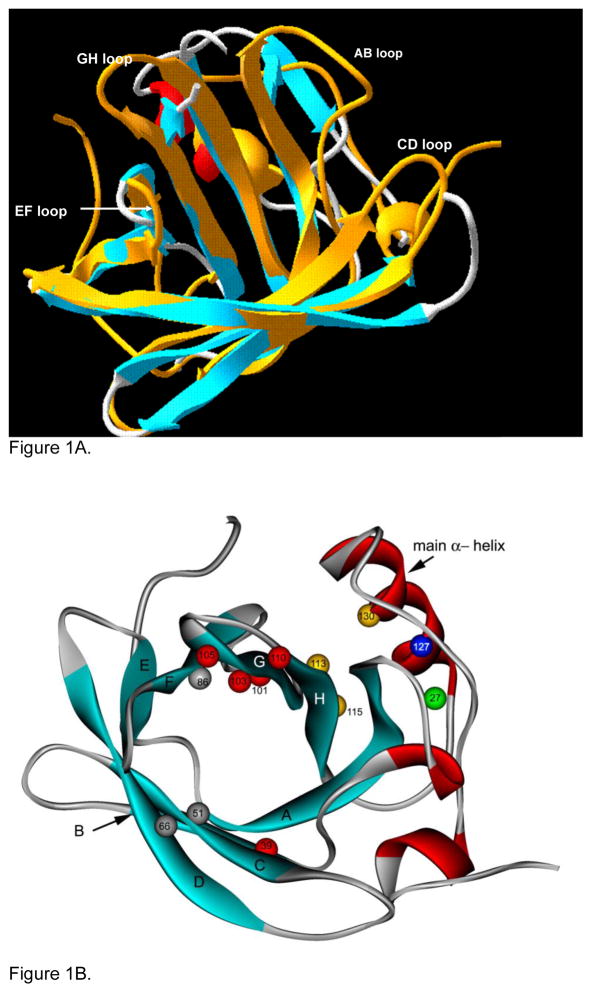
Figure 1.
A. Solution structure of tear lipocalin showing the positions of the 4 flexible loops (Gasymov et al. 2001 Biochemistry 40, 14754–14762) is superimposed on the crystal structures of tear lipocalin without the loops (adapted from Breusted et al. 2005 J. Biol Chem 280, 484–493). B. Cartoon structure of tear lipocalin illustrates the positions of the anti-parallel β strands (aqua ribbons), α-helical segments (red ribbons), loops (thin grey segments). The five amino acids with the greatest static quenching constants (indicative of strong ligand binding) for fatty acids are shown in red. Other key amino acid positions are discussed in text (numbered and colored balls). Adapted from solution structure (Gasymov et al., 2001 Biochemistry 40, 14754e14762).

