
Figure 2.
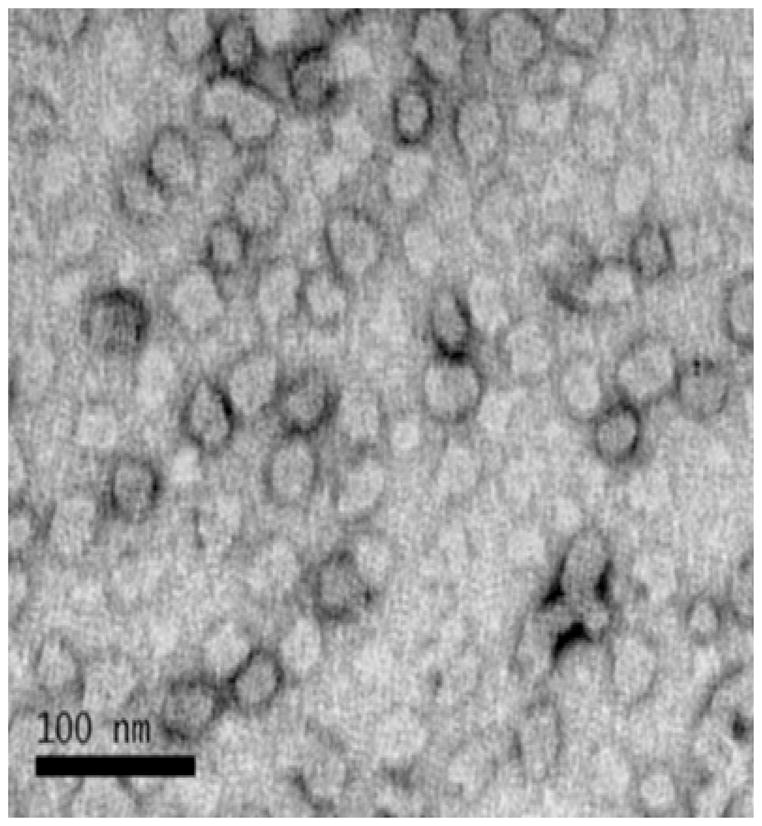
Transmission electron microscopy image of micelles formed from an aqueous solution of the diblock copolymer. A 0.5 mg/ml solution of the diblock copolymer in PBS was applied to a carbon coated copper grid for 30 minutes. The grid was fixed in Karnovsky’s solution and washed in cacodylate buffer once and then in water 8 times. The grid was stained with a 6% solution of uranyl acetate for 15 minutes and then dried until analysis. Transmission electron microscopy was carried out on a JEOL 1230 microscope.