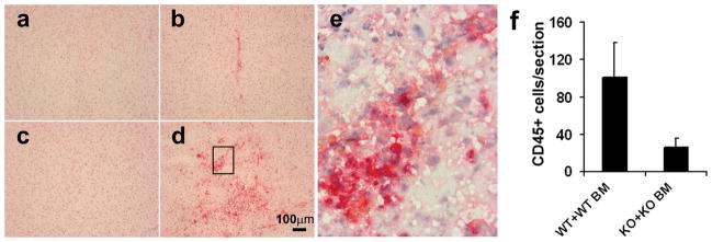
Figure 3. Homing of CD45+ cells to VEGF-induced mouse brain angiogenic foci was impaired in MMP-9 KO mice.
Representative images of immunohistochemical-stained mouse brain sections from WT+WT BM mice (c, d) and MMP-9 KO+KO BM mice (a, b) Many CD45+ cells (red) were detected around the AAV-VEGF injection site in the WT+WT BM brain (d), a few in the MMP-9 KO+KO BM brain (b), none in the AAV-LacZ-injected WT+WT BM brain (c) or KO+KO BM brain (a). High magnification photo (e) shows the boxed region in d. Bar graph shows the quantification of CD45+ cells around the injection sites (f), Data are mean±SD. N=4 per group.

