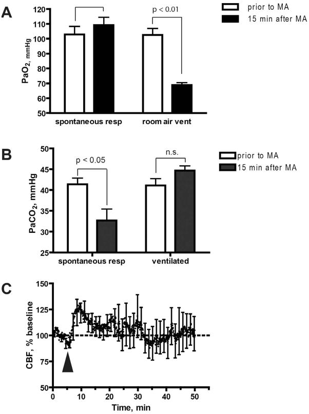
Figure 3. Mechanical ventilation prevents CBF decrease in response to MA.
A. Additional O2 is needed to provide adequate oxygenation in artificially ventilated mice. Oxygenation of arterial blood was within normal range before and after exposure to MA when mice were allowed to breathe spontaneously (note that RR reached 300 bpm after exposure to MA). In contrast, exposure to MA when mice were artificially ventilated with room air at 150 bpm resulted in a significant decrease in PaO2, relative to baseline levels (p = 0.0057, two-tailed t-test). Therefore, to avoid this non-physiological hypoxia, mice were ventilated in subsequent experiments with air containing 50% oxygen. B. Mechanical ventilation prevents the MA-mediated decrease in PaCO2. PaCO2 was measured prior to MA injection or 15 min after MA injection. Results represent mean ± SEM from 7 mice (not ventilated) and 5 mice (ventilated). The asterisk denotes that pCO2 in spontaneously breathing animals was significantly reduced at 15 min following exposure to MA, as compared to baseline (p = 0.017, two-tailed t-test). C. Mechanical ventilation prevents the MA-mediated decrease in CBF. CBF was measured in mechanically ventilated mice. Value are expressed as a percentage of baseline CBF, and represent mean ± SEM from 5 mice. Arrowheads indicate MA injection.

