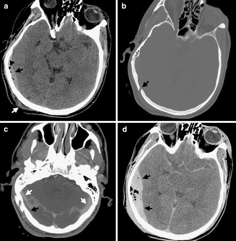
FIG. 2.
Venous epidural hematoma. (a) This patient sustained a blow to the back of his right occiput (coup site), as indicated by scalp soft tissue swelling (white arrow). Underlying the coup site is a lentiform EDH (black arrow). Several foci of pneumocephalus are noted (arrowhead) that indicate an associated skull fracture. (b) The displaced skull fracture (black arrow) is best seen on bone windows. (c) Contrast-enhanced CT venogram was obtained as the fracture line extended over the expected location of the right transverse sinus. The opacified transverse sinuses (white arrows) are patent, but the right transverse sinus is compressed and displaced from the inner table of the skull by the EDH (black arrow) caused by injury to the transverse sinus. Note that EDHs form superficial to the periosteal dural layer vesting the outer margin of the venous sinus, thereby possibly displacing the venous sinus away from the calvarium. (d) Follow-up CT 3 h after presentation shows substantial enlargement of the EDH (black arrows), which is less commonly seen with venous EDHs as compared to arterial EDHs. This was subsequently surgically evacuated. (High resolution version of this image is available in the electronic supplementary material.)

