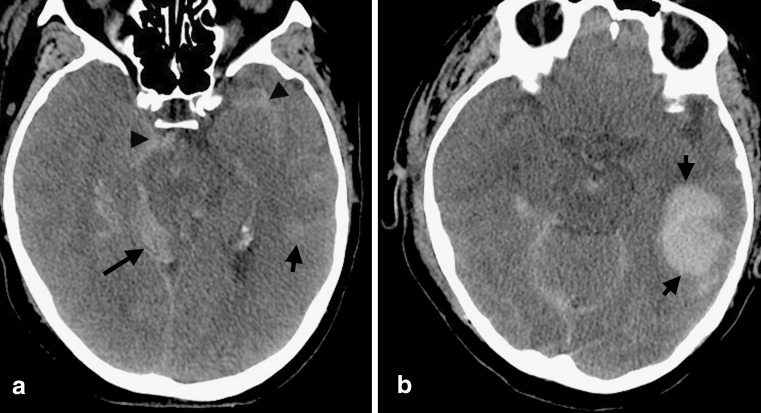
FIG. 7.
Blossoming of hemorrhagic contusions. (a) Multiple intracranial hemorrhages are seen in this patient, including a small, subtle left temporal hemorrhagic contusion (short arrow), SDH along the right tentorium (long arrow), and SAH in the basilar cisterns and Sylvian fissure (arrowheads). (b) Follow-up CT scan 6 h later demonstrates significant expansion of the left temporal contusion (short arrows) into an intraparenchymal hematoma, underscoring the importance of serial CT monitoring. (High resolution version of this image is available in the electronic supplementary material.)

