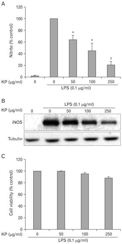
Figure 1.
Effect of KP on the NO secretion and iNOS expression in TG-elicited mouse peritoneal macrophages. (A) Cells were incubated with various concentrations of KP for 1 h and then stimulated with 0.1µg/ml LPS for 24 h at 37℃. The amount of nitrite released was measured by the method of Griess. Values are means±S.E. of three independent experiments. *p<0.05 and †p<0.01 vs LPS-treated group. (B) Cells were treated with KP and/or LPS as mentioned above and equal cytosolic extracts were analyzed by Western blotting with anti-iNOS antibody. Western blot detection of β-tubulin was estimated protein-loading control for each lane. (C) Cells were incubated with various concentration of UR in presence of 0.1µg/ml of LPS for 24 h. Then cell viability was measured by MTT assay as described in Materials and Methods. Data represent the relative viability to control group and are expressed as the means±S.E. of three independent experiments.

