Figure 3.
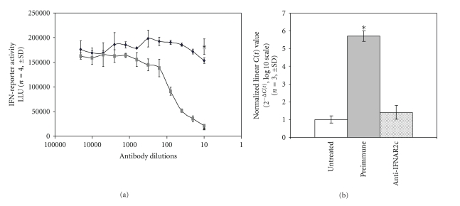
IFN-β induction of RGS1 is dependent on IFN-receptor activation. Neutralizing antisera recognizing the IFNAR2c IFN-receptor was incubated with PBMCs at various concentrations to determine the dilution necessary to achieve greater than 95% inhibition of an IFN-reporter assay (see Section 2). Figure 3(a): Cells (HT1080LUC) were incubated as previously described with preimmune (dark squares) or IFNAR2c antisera (gray squares). Antibody dilution is represented on the x-axis. The extent of IFN-reporter activation is shown on the y-axis represented as luciferase light units (LLU). IFN-reporter activity for IFN-β-stimulated HT1080LUC cells in the absence of antisera is also shown for comparison (single gray square). Data are presented as mean (n = 4) ±SD. Figure 3(b): TaqMan analysis of IFN-β induced expression of RGS1 in the absence (untreated and preimmune) or presence (anti-IFNAR2c) of an IFN-receptor neutralizing antisera. RGS1 induction is expressed as linearized C(t) values normalized to GAPDH (n = 3, mean ± SD). *P < .05.

