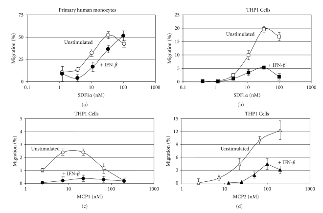
Figure 7.
Effect of IFN-β on chemokine-dependent migration of monocytes. Monocyte transmigration across an artificial barrier in response to chemokine stimulation was determined as described in Section 2. Primary monocytes incubated with IFN-β (1000 IU/107 cells) for 20 h were stimulated with various concentrations of SDF1α (Figure 7(a)). IFN-β-stimulated (dark circles) and unstimulated (open circles) cells are shown. The concentration of SDF1α (nM SDF1α) is shown on the x-axis along with transmigration, as percent (% migration) of cells applied, on the y-axis. The monocytic cell line THP-1 was incubated with IFN-β (1000 IU/107 cells) for 20 hours and then stimulated with increasing concentrations of either SDF1α (Figure 7(b)), MCP1 (Figure 7(c)), or MCP2 (Figure 7(d)). The concentrations of SDF1α, MCP1, and MCP2 are shown on the x-axis (nM chemokine) and results are presented as percent of total cell migration (% migration). Data represents mean ± SD of n = 4.

