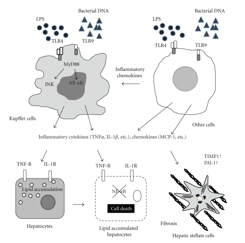
Figure 1.
TLRs and downstream signaling in NAFLD. Kupffer cells respond to TLR ligands such as LPS and bacterial DNA through TLR4 and TLR9, respectively. Upon TLR ligation, MyD88, an adaptor molecule, is recruited to transmit the signals that activate NF-κB and JNK. Activated Kupffer cells produce inflammatory cytokines such as TNFα and IL-1β and chemokines such as MCP-1 (CCL2). These inflammatory cytokines and chemokines induce lipid accumulation in hepatocytes and cell death. In addition, TNFα and IL-1β promote liver fibrosis by activating hepatic stellate cells. Other cells including hepatic resident cells, infiltrated cells into the liver, and adipose tissue macrophages produce various mediators in response to TLR ligands. These pathways also contribute to the development of NAFLD.

